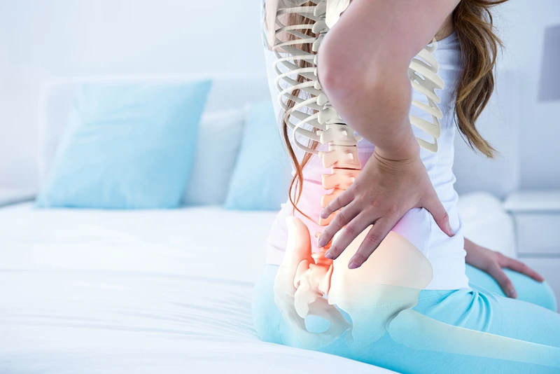
Sciatica is a common name for what doctors call “lumbar radiculopathy” — in simple terms, irritation of a nerve in the lower back (often referred to as a “pinched nerve”). In most cases, this happens because a spinal disc bulges or herniates and irritates a nearby nerve. Age-related wear and tear, narrowing of the spinal canal, or slight shifting of one vertebra on another can also play a role.
When this nerve becomes irritated, the pain doesn’t just stay in the back — it often travels down the leg, sometimes all the way to the foot. People commonly describe it as sharp, burning, shooting, or even electric. There may also be pins and needles, numbness, or a feeling of weakness in the leg.
Symptoms often feel worse when sitting for long periods, bending forward, or even when coughing or sneezing, as these movements increase pressure in the lower back. Many people find relief lying on their back with their knees supported. During a physiotherapy assessment, we check your strength, sensation, reflexes, and specific nerve tests to work out whether a nerve is truly involved.
Current international guidelines — including those from the National Institute for Health and Care Excellence (NICE, 2020 updated guidance) and the North American Spine Society (NASS, 2022 guideline update) — recommend that most people with sciatica are managed conservatively (without surgery) provided there are no red flags (such as worsening neurological deficit or bowel/bladder dysfunction suggestive of cauda equina syndrome). Routine early imaging is not recommended unless serious pathology is suspected or symptoms persist beyond 6–8 weeks.
Although we often think of this nerve pain as simply the result of a disc pressing on a nerve, research shows that chemical and inflammatory factors play a major role too. When disc material leaks out, it doesn’t just physically contact the nerve — it releases inflammatory substances that irritate nerve fibres and surrounding tissues. These chemicals can sensitize the nerve and trigger pain signals even when there isn’t significant mechanical compression. This helps explain why some people have severe leg pain despite only a small herniation seen on MRI, or why pain can improve as inflammation settles even if the disc herniation is large and hasn’t shrunk much on scans. This chemical component of nerve pain is increasingly recognised in modern pain science, and is part of why treatments like physiotherapy can be effective even without major structural change.
Reassuringly, high-quality research shows that the majority of these “pinched nerves” improve within 6–12 weeks without surgery. Recent evidence also emphasises that disc herniations frequently reduce in size naturally over time due to inflammatory resorption processes. A recent review suggests that up to around 60–70% of lumbar disc herniations show radiological resorption. Interestingly though, clinical improvement in pain and function does not depend on this reabsorption. Studies have demonstrated that patients may experience substantial pain relief and nerve symptom resolution before there is measurable reduction in herniation size on MRI. This is because factors such as reduction of chemical inflammation around the nerve root, decreased nerve irritation, and central modulation of pain pathways — contribute significantly to symptom improvement. For example, one clinical series reported that patients’ symptoms often improved within weeks, while radiological regression took several months or more to become apparent, highlighting that functional recovery can precede or occur independently of measurable anatomical change. It’s all good news!
Physiotherapy plays an important role in recovery — not by “pushing discs back in,” but by helping the irritated nerve settle, restoring movement, and gradually building strength and confidence again.
In the early stages, treatment focuses on calming pain and reducing nerve sensitivity. This may include education (which research shows reduces fear and improves outcomes), guidance on positions that ease pressure on the nerve, gentle movement, and specific exercises tailored to your symptoms.
Physiotherapists often use manual therapy – spinal mobilisation, massage and neural mobilisation – to help reduce pain and improve movement. This is always combined with active rehabilitation, as exercise-based approaches show the strongest evidence for long-term benefit. Staying active, within reason, has been shown to support recovery better than prolonged bed rest, which actually does more harm than good.
As symptoms begin to settle, rehabilitation shifts toward restoring normal movement in the spine and hips, improving core and trunk strength, and addressing any contributing factors such as stiffness, weakness, or poor movement patterns. Strengthening and graded exercise programmes are strongly supported in the research for improving function and reducing long-term recurrence.
Importantly, physiotherapy also helps you understand what is happening in your body. Research consistently shows that people who understand their pain, remain active, and gradually return to normal activity tend to recover better than those who avoid movement out of fear.
Every case is different, but with the right guidance, most people can return to normal activities without the need for injections or surgery.
Juli-Ann Riley, Riley Physiotherapists